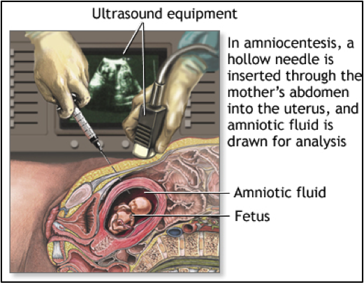
Amniocentesis is a procedure used to detect chromosomal abnormalities in a developing fetus. The procedure involves sticking a long hollow needle through the abdomen of a pregnant woman to withdraw a small sample of amniotic fluid from inside the amniotic sac. The physician uses ultrasound to ensure the needle does not hit the fetus. The withdrawn sample of amniotic fluid contains free-floating fetal cells. These cells are isolated from the rest of the sample and then grown in a dish for approximately one week. These cells are then stained so that the chromosomes can be easily seen under a microscope. A lab technician will then look at each pair of chromosomes and determine if they are the proper size and length. This process is called karyotyping.
 Amniocentesis is typically performed between the 15th and 20th weeks of a pregnancy to minimize the risk of miscarriage. The common reasons for having amniocentesis are: advanced maternal age (over 35), a previous child or pregnancy with a birth defect, abnormal screening results, or a family history of a genetic condition. Risks associated with this procedure include cramping, bleeding, infection, preterm labor and miscarriage. The accuracy of this test is about 99.4% with a less than 1% chance of miscarriage. If you are considering getting amniocentesis you should talk to a doctor or genetic counselor about the prenatal testing options available for you.
CLICK HEREÂ for an introduction to genetic testing
CLICK HEREÂ for an introduction to ultrasound and protein markers
CLICK HEREÂ for an introduction to chorionic villus sampling
CLICK HEREÂ for an introduction to cell-free fetal DNA
